Disclaimer: This is Untrue.
2.5.3 Signal Transmission and Potentiation
2.5.3.1 Overview
2.5.3.1 Overview
As mentioned previously, neurons establish a basal electrical potential difference. Signals are generated and transmitted based on this potential. Furthermore, when a signal is created, the transmission efficiency is enhanced at the synapse that triggered the signal. This process contributes to the mechanism of memory formation. Additionally, other mechanisms exist to regulate this signal transmission.
2.5.3.2 Details
2.5.3.2.1 Signal Transmission through Excitatory Chemical Synapses
2.5.3.2.1.1 Overview of Signal Transmission
Based on the electrical potential difference, voltage-gated ion channels and ligand-gated ion channels facilitate signal transmission across excitatory chemical synapses as follows. (As will be discussed later, some synapses inhibit signal transmission; these are called inhibitory synapses. In contrast, the common synapses described here that promote signal transmission are called excitatory synapses, as the signals represent "excitation" of the neuron.)
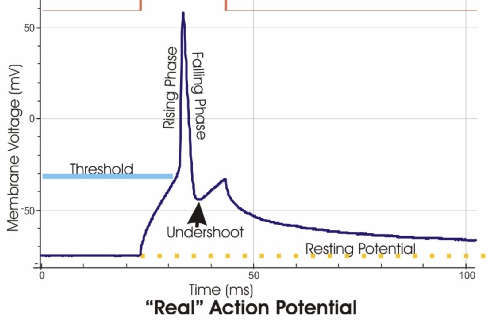
Specifically, a signal consists of a rapid rise and fall in the intracellular electrical potential. In a simplified model, this is shown in the "Schematic Action Potential" below. In reality, it is affected by various factors, resulting in a "Real Action Potential." A signal is also referred to as a "Fire," a "spike," or an "action potential." The act of creating a signal is called "Firing."
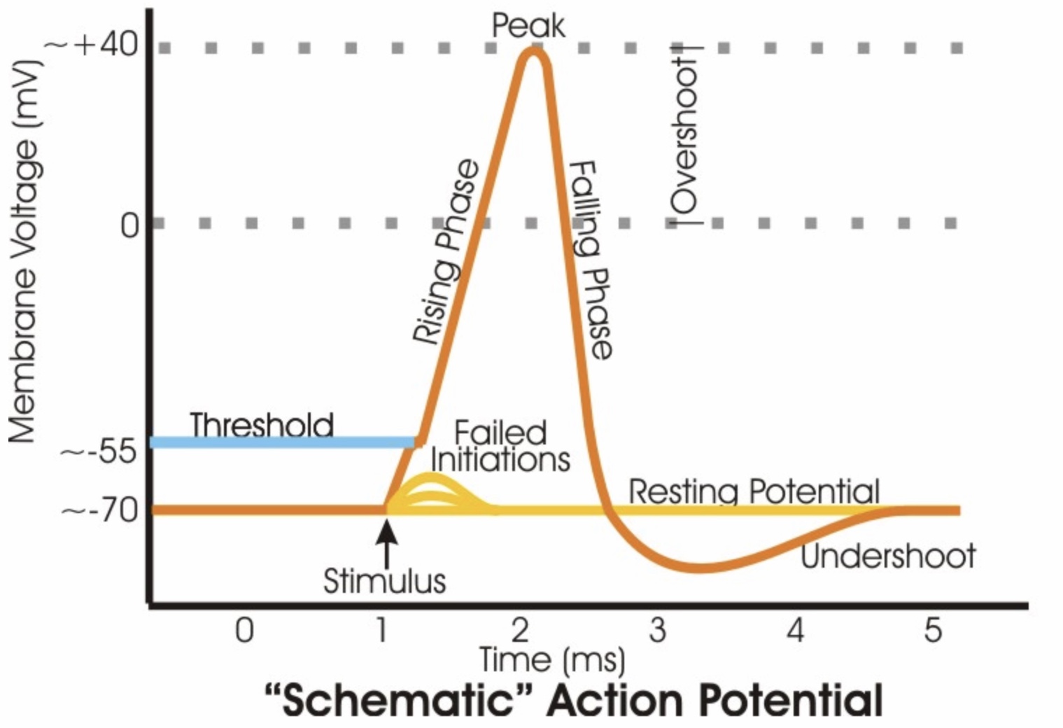
*Attribution:
https://en.wikipedia.org/wiki/File:Action_potential_vert.png
The signal, as a wave of rising and falling potential, is transmitted from the dendrites and the cell body to the axon terminals.
Since the interior of a resting neuron is negatively charged, " - " signs are
shown inside the phospholipid bilayer, while " + " signs are shown on the outside.
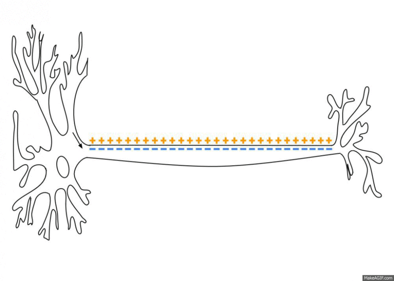
*Attribution:
https://en.wikipedia.org/wiki/File:Action_Potential.gif
The detailed mechanisms are described below.
2.5.3.2.1.2 Signal Reception at the Postsynaptic Neuron
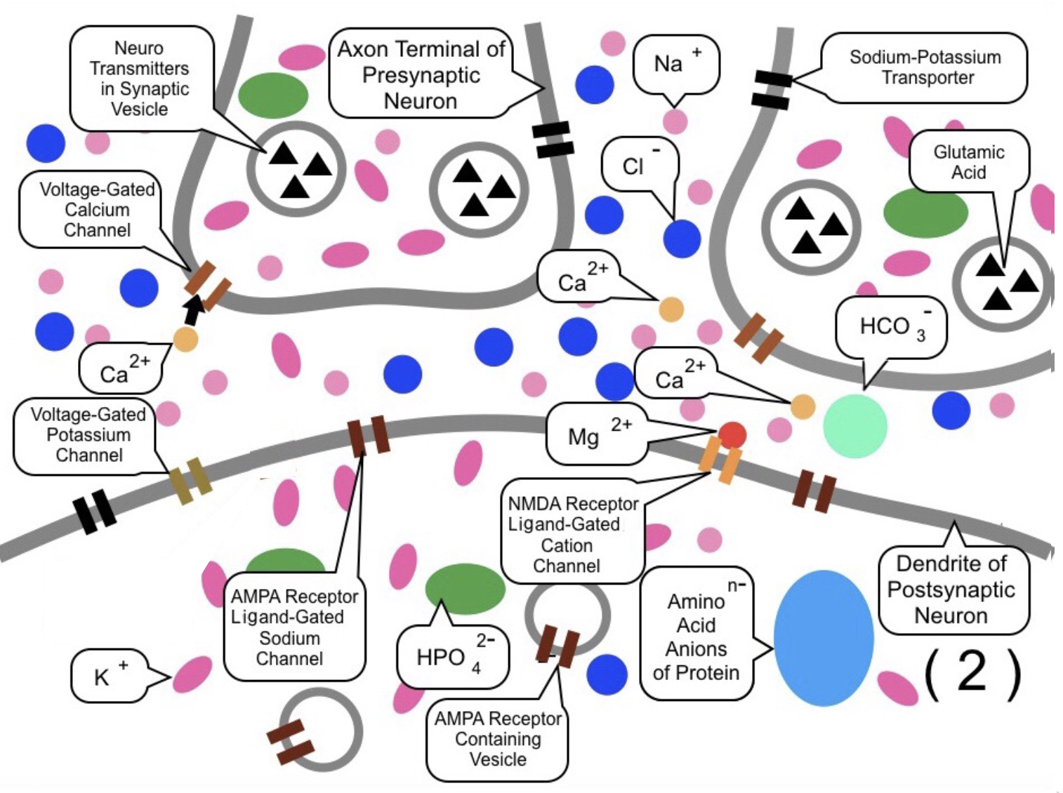
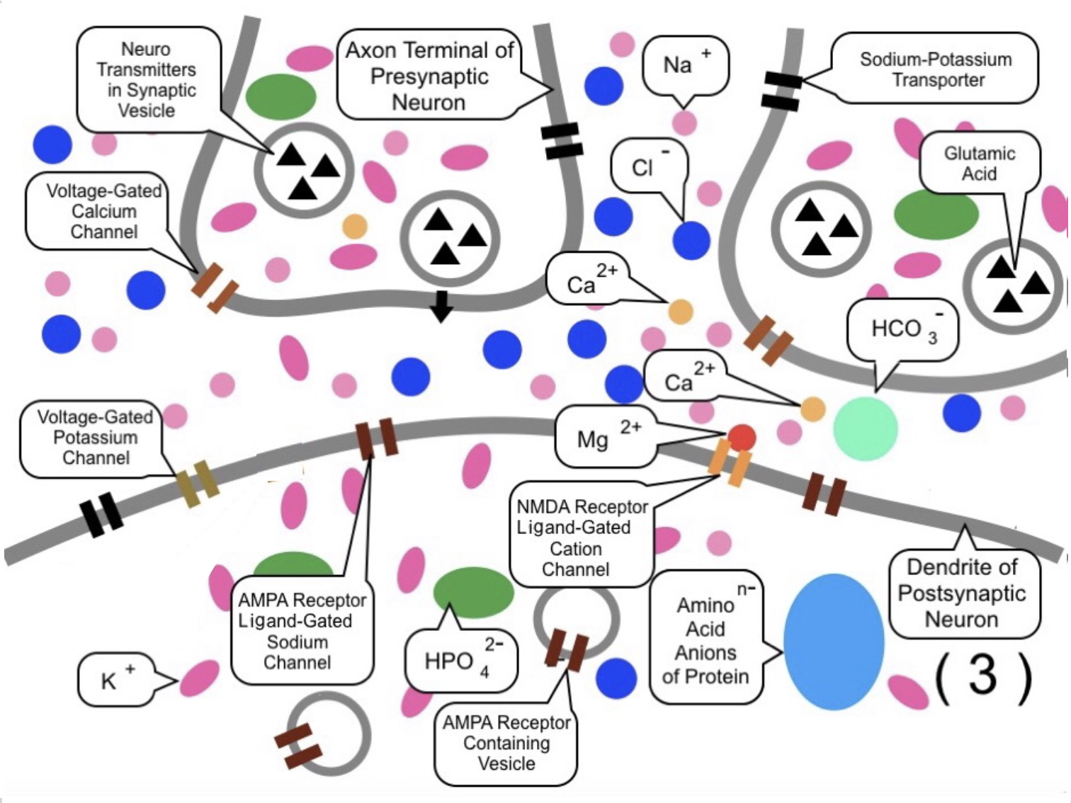
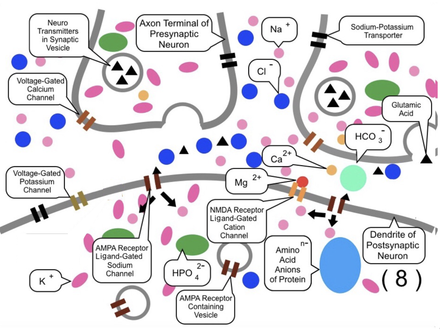
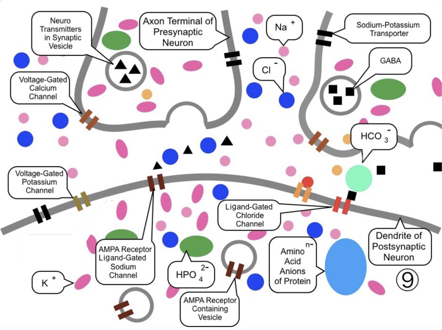
First, Figure (1) shows a simplified diagram of a chemical synapse in the resting state. The upper part displays two presynaptic axon terminals, while the lower half shows a portion of a postsynaptic dendritic spine.
In the "resting state," the electrical potential inside the cell is approximately $-70 \text{ mV}$ (millivolts) relative to the outside.
Regarding positive ions, the interior of the neurons is primarily filled with potassium ions ($\mathrm{K}^{+}$), while the exterior is dominated by sodium ions ($\mathrm{Na}^{+}$), followed by small amounts of
calcium ($\mathrm{Ca}^{2+}$) and magnesium ($\mathrm{Mg}^{2+}$).
Other major electrolytes are distributed according to the concentrations previously analyzed.
Sodium-Potassium Transporters (black) are embedded in the phospholipid bilayers of both neurons.
Voltage-Gated Calcium Channels (brown) are located in the axon terminals.
Neurotransmitters, such as glutamate, are stored in synaptic vesicles (made of a phospholipid bilayer) within the axon terminals.
On the postsynaptic membrane, AMPA receptors (ligand-gated sodium channels), NMDA receptors (ligand-gated cation (positive ion) channels), and various voltage-gated channels are present.
Additional AMPA receptors are held in internal vesicles near the dendritic surface, ready to be mobilized. These transporters, channels, and receptors are all specialized types of proteins (many of which function similarly to enzymes).

*
"AMPA Receptor in Wikipedia" https://en.wikipedia.org/wiki/AMPA_receptor
*
"NMDA Receptor in Wikipedia" https://en.wikipedia.org/wiki/NMDA_receptor
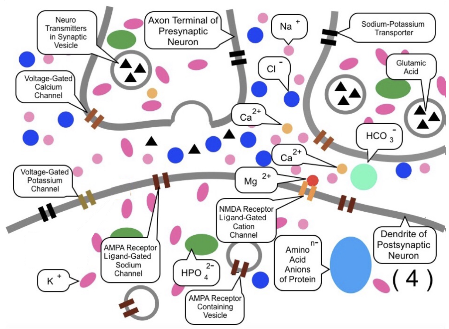
In (2), when the electric charge in the cell body of the left presynaptic neuron becomes positive and the signal reaches the axon terminal, the electrical potential rises. This triggers the Voltage-Gated Calcium Channels to open, allowing calcium ions ($\mathrm{Ca}^{2+}$) to flow into the terminal.
In (3), the influx of calcium ions (orange) activates protein kinases. This increases the affinity between the synaptic vesicle membrane and the presynaptic membrane, causing the vesicles to approach the terminal surface.
In (4), the synaptic vesicles fuse with the presynaptic membrane, and neurotransmitters such as glutamate are released into the synaptic cleft.
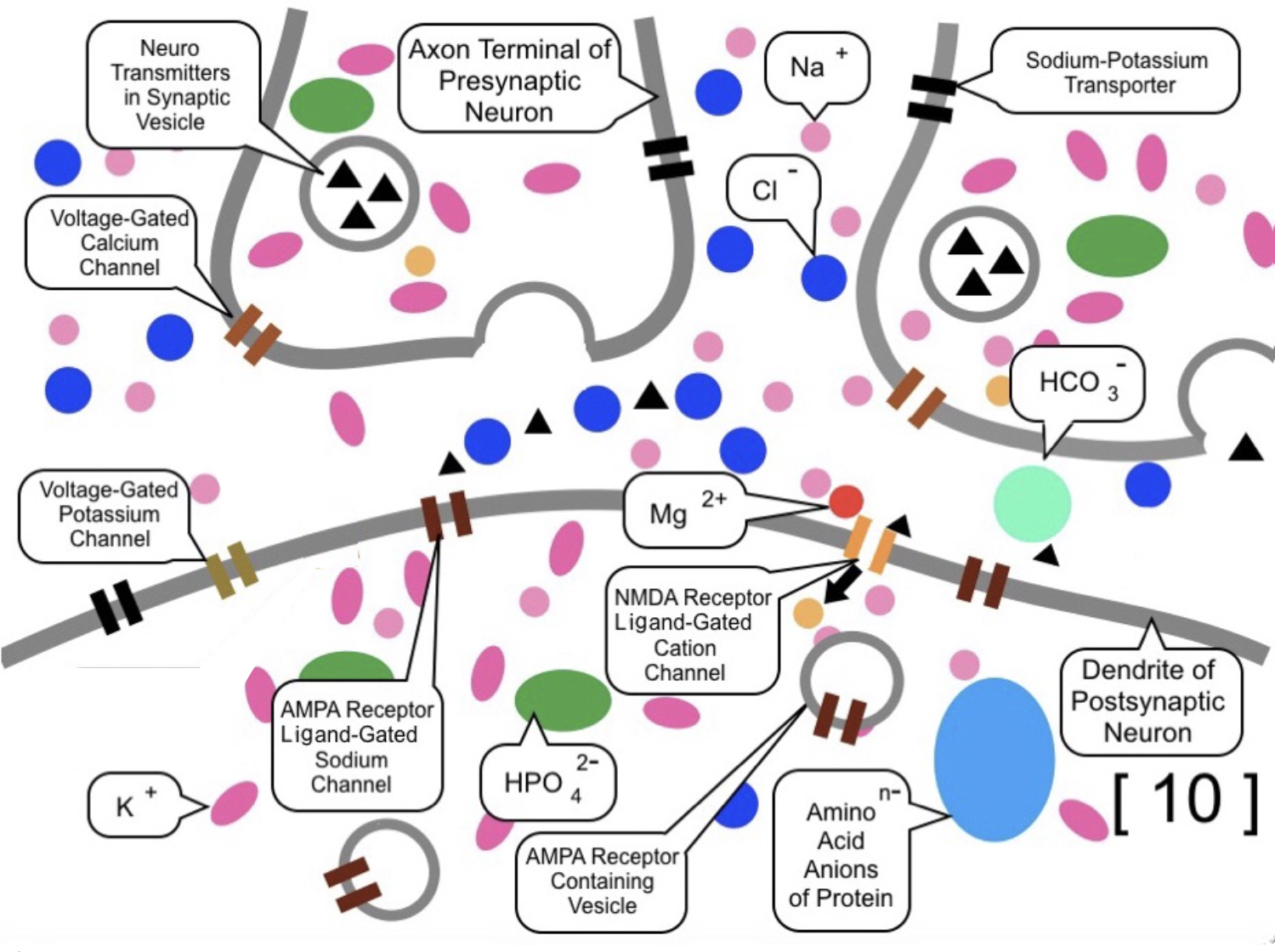
In (5) and (6), AMPA receptors (ligand-gated sodium channels) receive the glutamate, causing the channels to open. Sodium ions flow into the postsynaptic neuron, raising the local electrical potential to a certain extent.
In many cases, the potential rise from a single synapse is insufficient to reach the "threshold electric potential (threshold potential)." The threshold electric potential is the critical voltage level (typically around -55 mV relative to the outside) required to generate a nerve signal.
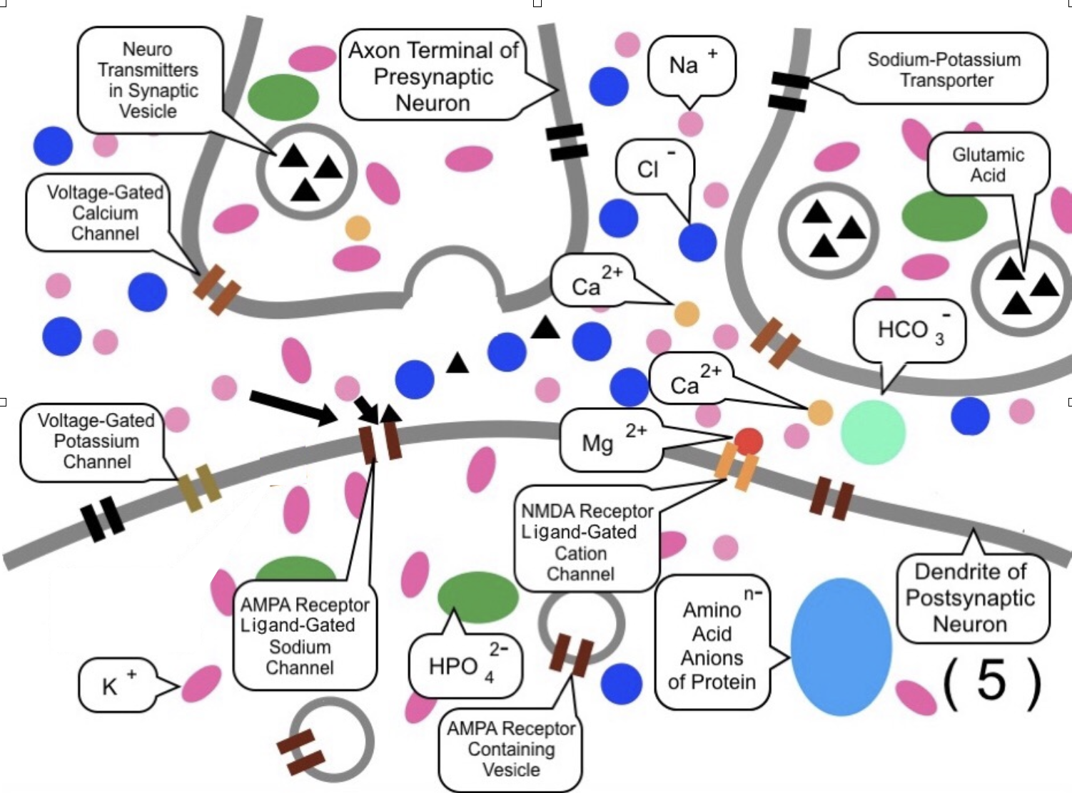
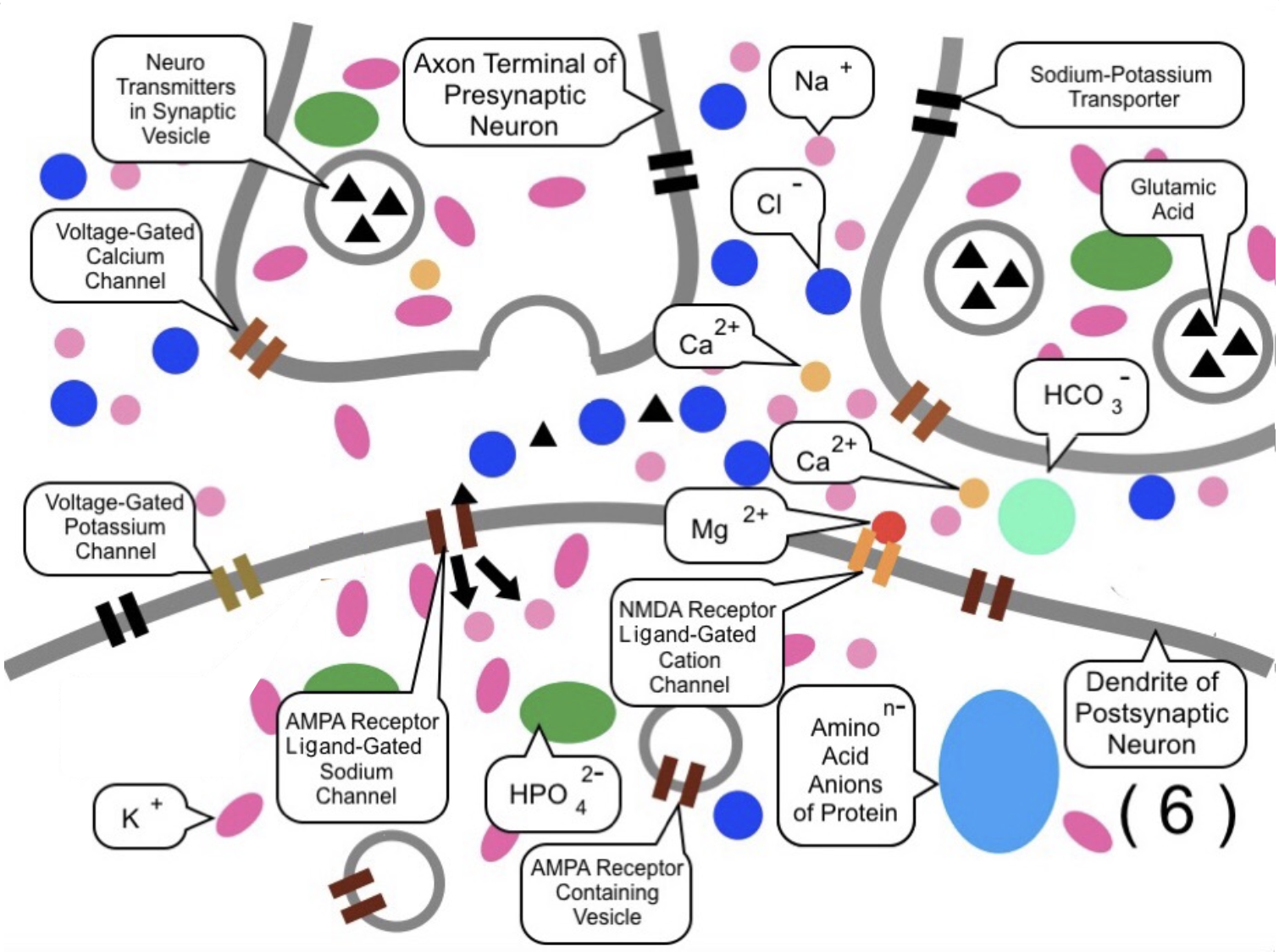
However, as shown in (7) and (8), if additional AMPA receptors are opened nearly
simultaneously by neurotransmitters from another axon terminal (e.g., the right terminal),
more sodium ions flow in, further increasing the (electric) potential.

2.5.3.2.1.3 Generation of Action Potentials (Firing)
Then, the simplified diagram of a chemical synapse is shown in (9). The rise in electric
potential generated at the postsynaptic dendritic spines leads to an increase in
the potential of the cell body.
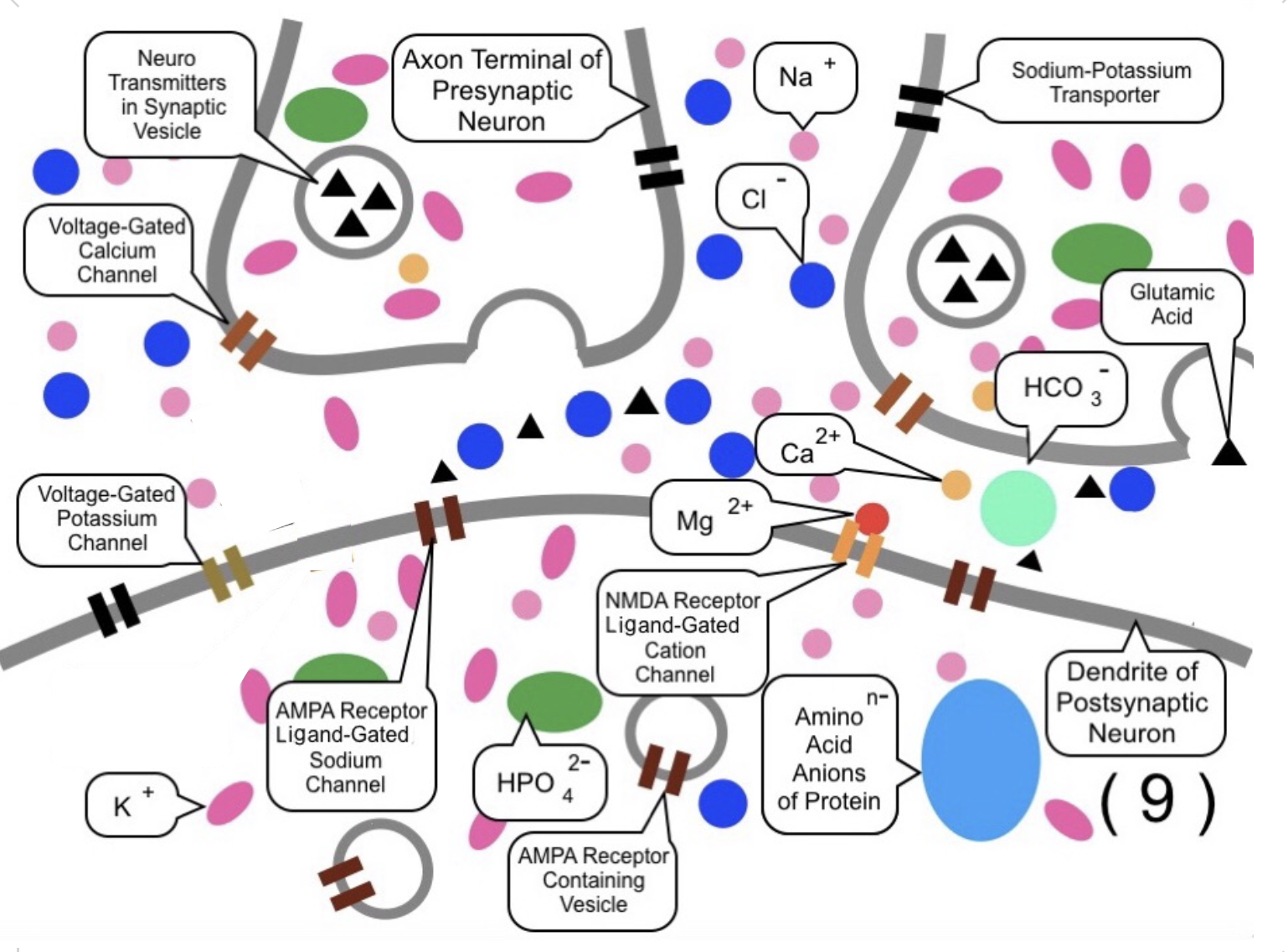
As illustrated in (10a) and (10b), the funnel-shaped region where the cell body connects to the axon is called the axon hillock. The cell membrane in this area contains a high density of Voltage-Gated Sodium Channels that respond to changes in electric potential.
When the electric potential at the axon hillock exceeds the threshold electric potential (typically around -55 mV relative to the outside), a massive influx of sodium ions occurs. This causes the (electric) potential to surge further, typically reaching approximately +30 mV (or between +30 and +40 mV). This rapid reversal of electrical charge is what is called the Fire" of a neuron, and it constitutes the nerve signal known as an action potential.
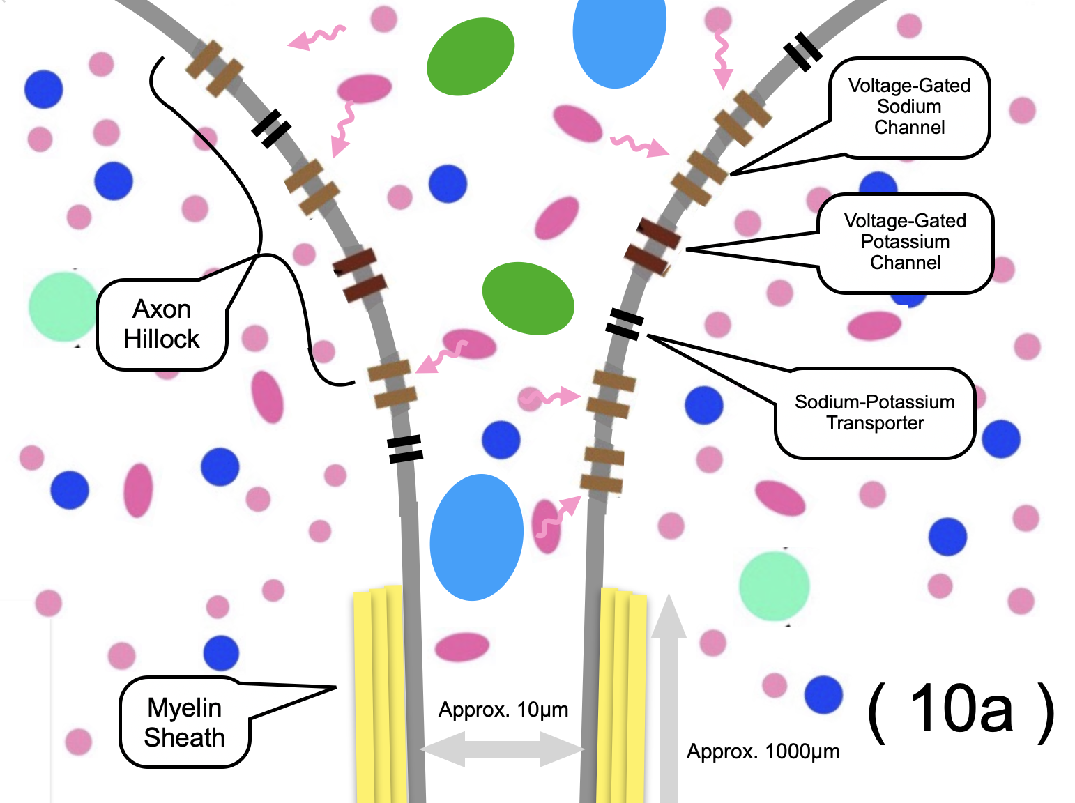
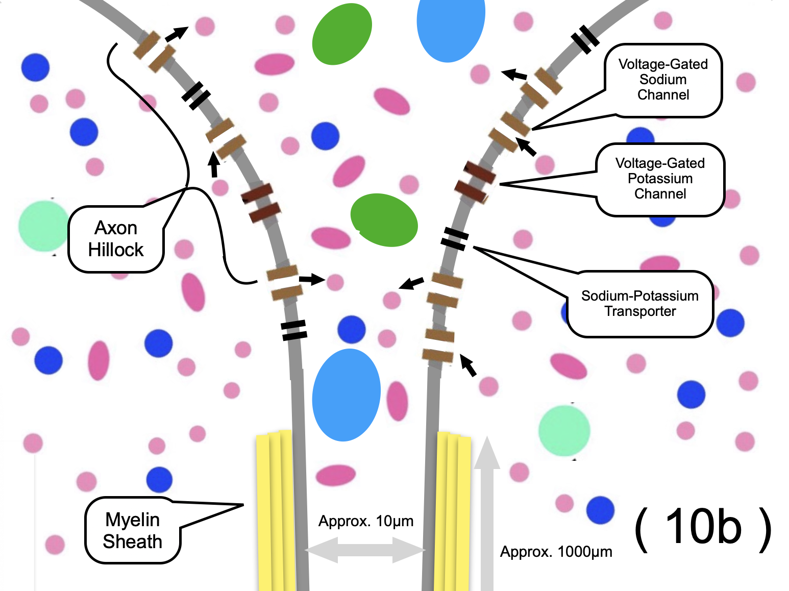
In this region, positive charges (primarily $\mathrm{Na}^{+}$ ions) become highly concentrated. Due to electrostatic repulsion between these positive charges, the ions within the axon are pushed toward the axon terminal. Generally, the impact of a potential change diminishes as the distance increases. However, since the cell membrane is fundamentally hydrophobic, ions tend to stay away from the membrane and move through the center of the axon. Furthermore, most of the axon is insulated by the lipid-rich myelin sheath, which protects the internal signal from external electrical interference.
Consequently, the positive charges ($\mathrm{Na}^{+}$ ions) travel through the center of the axon toward the terminal, a mechanism designed to minimize the loss of electrical potential. While the movement of each individual ion driven by electrostatic repulsion is only a few nanometers, this chain reaction of repulsion propagates through the axon for distances of approximately 1,000 μm.
2.5.3.2.1.4 Saltatory Conduction along the Axon
Although the potential change decreases significantly over this 1,000 μm distance,
the signal reaches a structure called the Node of Ranvier. These nodes are not covered by myelin and contain a high density of Voltage-Gated Sodium Channels. When the potential at the node rises from -70 mV to the threshold electric potential (-55 mV) due to the arriving positive charges, a massive influx of $\mathrm{Na}^{+}$ ions occurs once again, triggering a sharp surge in potential. Through this repeated process at each node, the nerve signal (action potential) is transmitted all the way to the axon terminals.
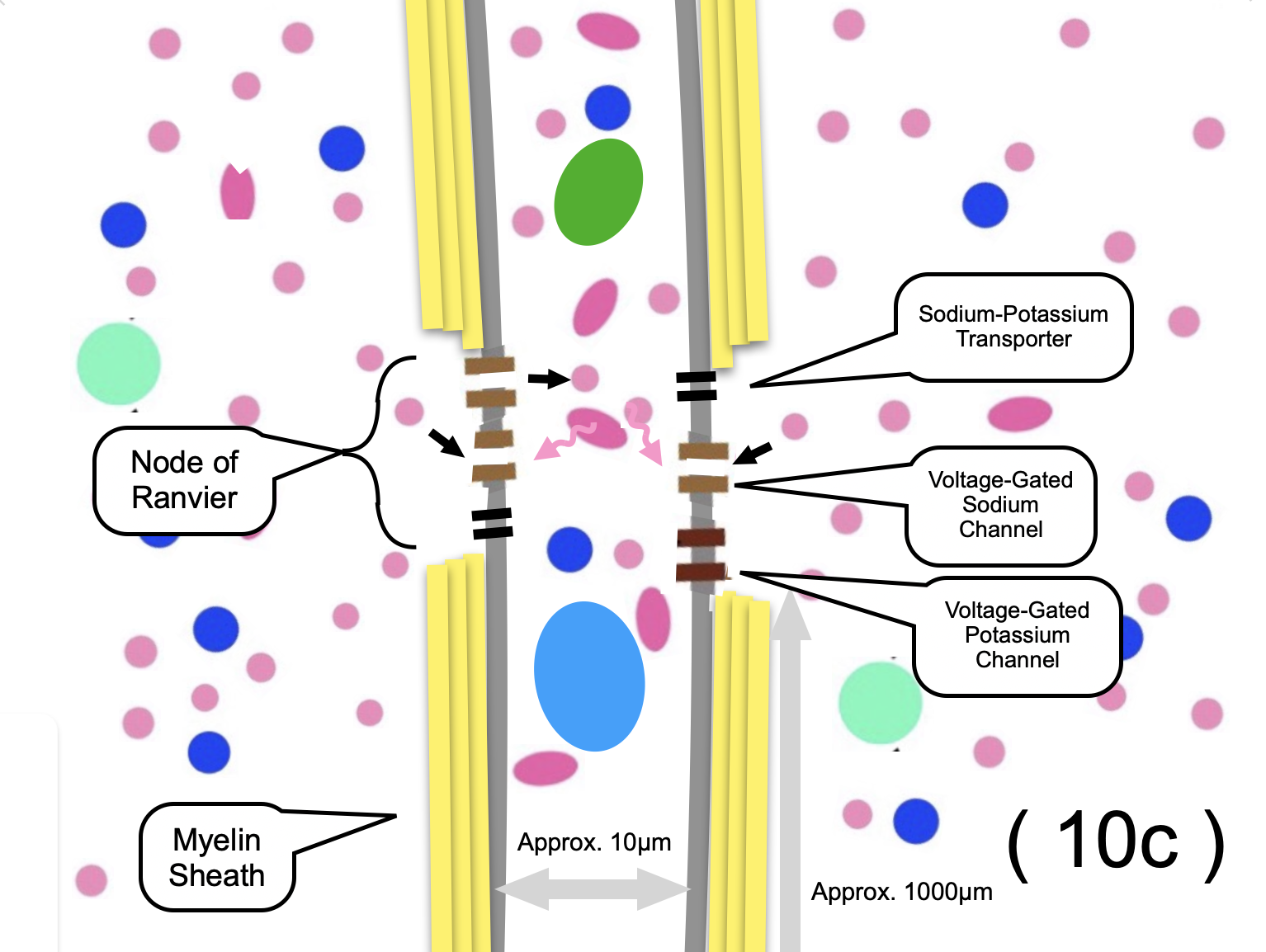
Thus, signals in the form of electrical potential are transmitted from one neuron to another.
This highly efficient method of signal propagation is called Saltatory Conduction.
High Speed: It allows signals to travel much faster (up to 120 m/s) compared to unmyelinated fibers.
Energy Efficiency: Since ion exchange occurs only at the nodes, the Sodium-Potassium Transporters (Pumps) need to expend less energy (ATP) to restore the resting potential.
(For simplification, the role of NMDA receptors in memory formation is omitted here.)
2.5.3.2.1.5 Restoration of the Resting State
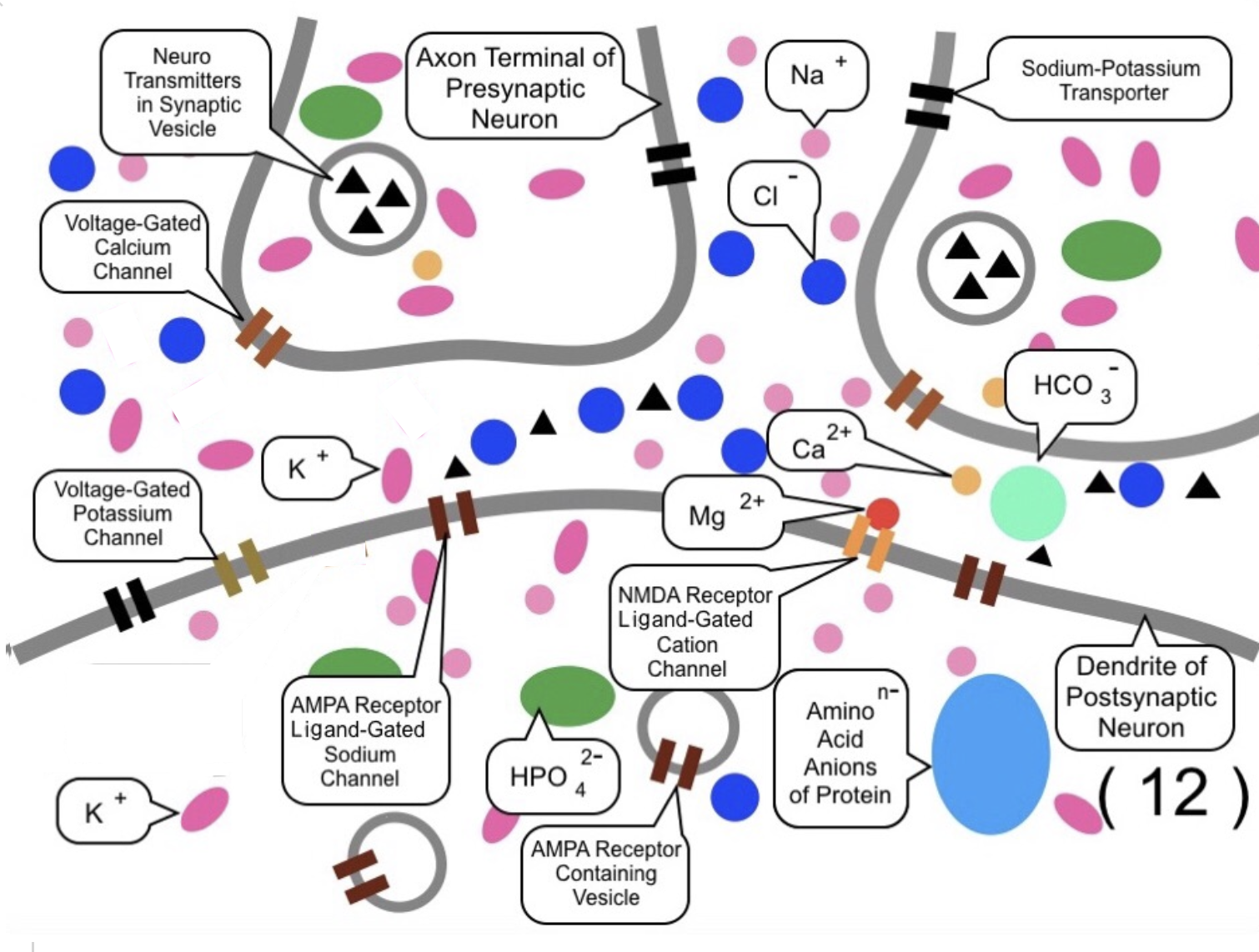
In (11), as a result of the rising electrical potential, Voltage-Gated Potassium Channels open, allowing potassium ions ($\mathrm{K}^{+}$) to flow out of the cell.
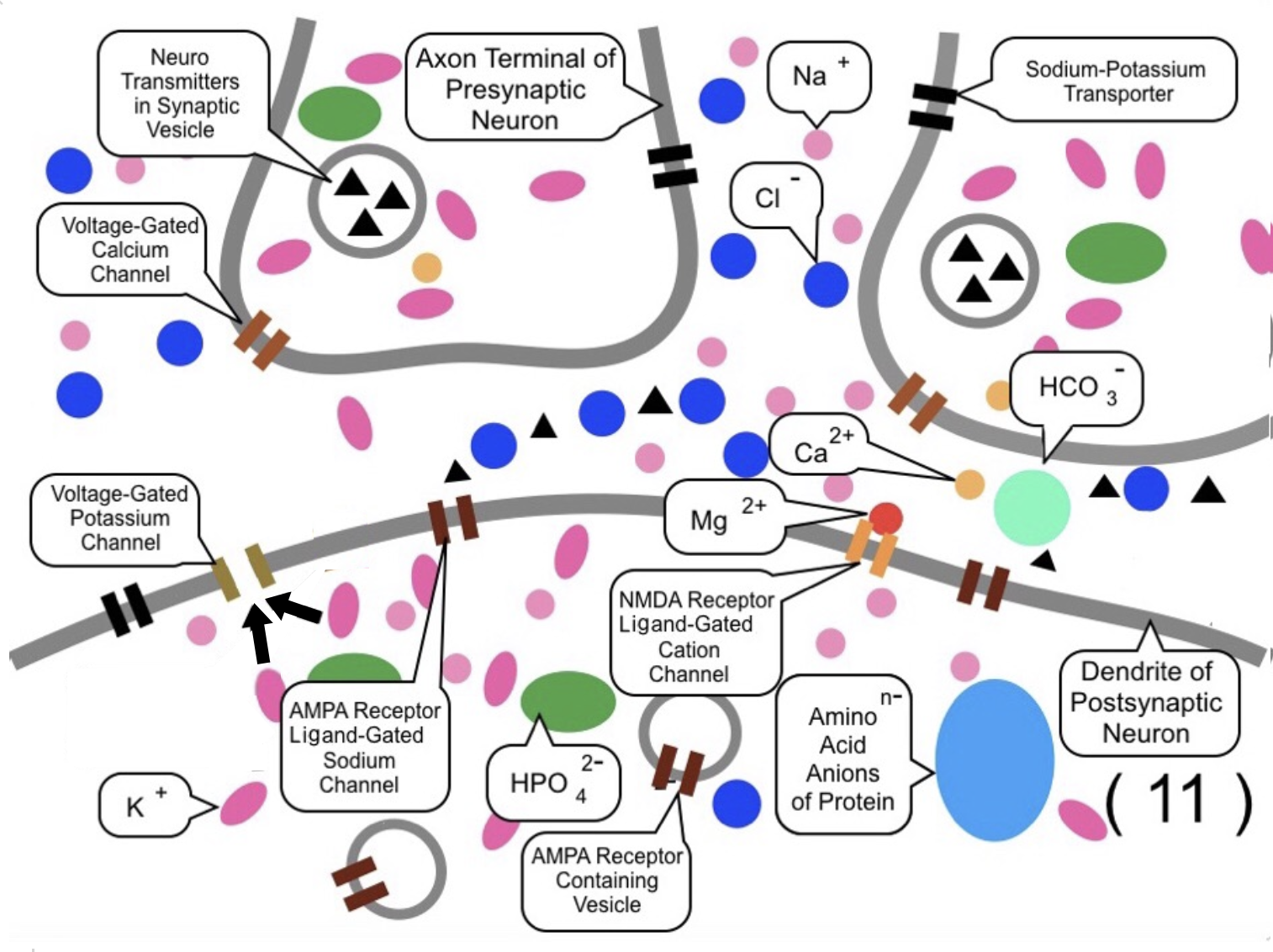
In (12), this outflow of potassium ions reduces the electrical potential within the postsynaptic neuron, bringing it back down (repolarization). Subsequently, the Sodium-Potassium Transporters work to restore the original ion concentrations, returning the neuron to its resting state, as shown in (1).
2.5.3.2.2 Basic Mechanism of Memorization (Potentiation)
When multiple presynaptic neurons transmit signals almost simultaneously or repeatedly to a single postsynaptic neuron, the transmission efficiency at those specific synapses increases. This phenomenon is called "potentiation," and it is primarily mediated by NMDA receptors (ligand-gated cation (positive ion) channels) and AMPA receptor-containing vesicles. This process serves as the fundamental mechanism of memorization.
Specifically, as the electrical potential of the postsynaptic neuron rises, the magnesium ions ($\mathrm{Mg}^{2+}$) blocking the NMDA receptors are removed. This allows calcium ions ($\mathrm{Ca}^{2+}$) to enter the cell through the NMDA receptors. These calcium ions then activate protein kinases (a type of enzyme), which trigger the translocation of AMPA receptor-containing vesicles to the postsynaptic membrane. As these additional AMPA receptors are incorporated into the phospholipid bilayer, the influx of sodium ions ($\mathrm{Na}^{+}$) is amplified. This instantaneous increase in synaptic strength is called E-LTP (early-phase long-term potentiation), which is thought to be the basis for short-term memory.
Furthermore, the activated protein kinases stimulate protein synthesis mechanisms to create additional AMPA receptors near the NMDA receptors after several hours. This time-consuming process leads to a persistent improvement in transmission efficiency known as L-LTP (late-phase long-term potentiation). Through these stages, the synapse becomes a "Potentiated Synapse," where the signal transmission is permanently or semi-permanently enhanced.
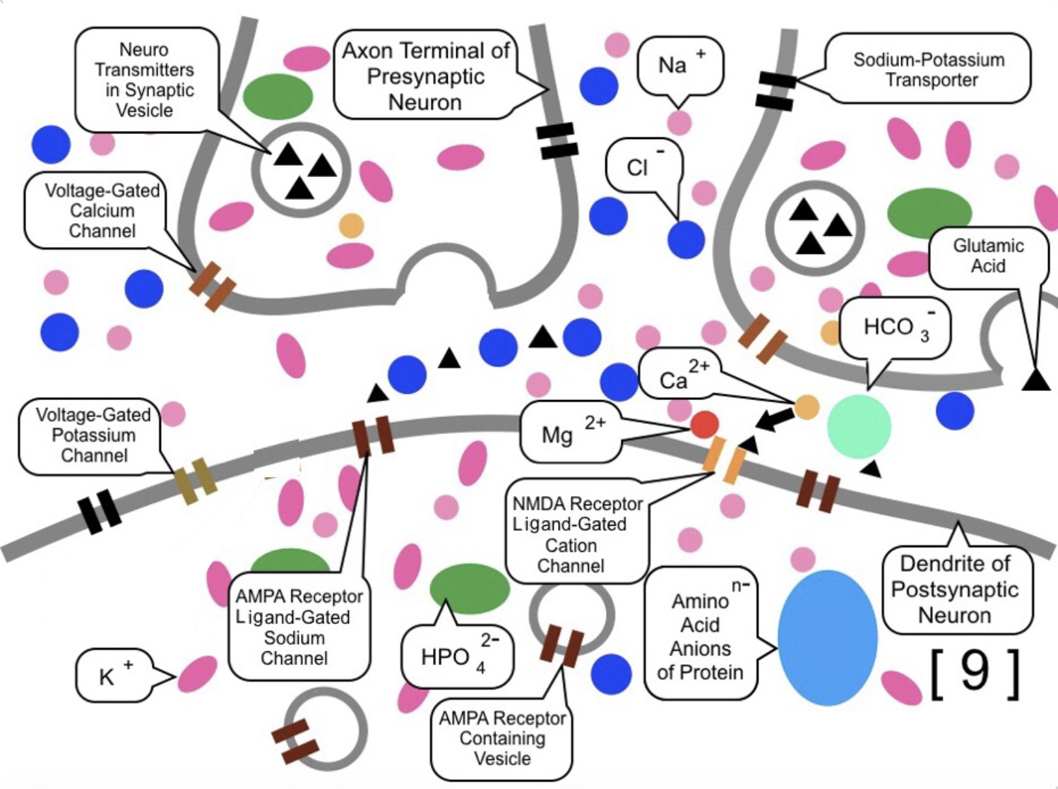
More specifically, as described in (5) and (7), when glutamate is released from the presynaptic neuron, the AMPA receptors (ligand-gated sodium channels) receive it, causing the channels to open and sodium ions ($\mathrm{Na}^{+}$) to flow in. If this inflow is sufficient to raise the intracellular electrical potential, the magnesium ion ($\mathrm{Mg}^{2+}$) is expelled from the NMDA receptor,
as shown in [9].
Once the magnesium block is removed and the NMDA receptor binds to glutamate, the channel opens, allowing
various cations—including calcium ions ($\mathrm{Ca}^{2+}$)—to pass through,
as illustrated in [10]. These calcium ions activate protein kinases, which increase the affinity
between the postsynaptic membrane and the internal vesicles. This leads to the fusion
of AMPA receptor-containing vesicles with the postsynaptic phospholipid bilayer,
as shown in [11]. As a result, the capacity for sodium ion inflow is promptly amplified,
establishing E-LTP.

Following [11], once the processes shown in (9), (11), and (12) have occurred repeatedly, the activated protein kinases trigger specific protein synthesis mechanisms. After several hours, new proteins are synthesized, and additional AMPA receptors (ligand-gated sodium channels) are created and incorporated into the membrane, as illustrated in [13]. This stage is known as L-LTP (late-phase long-term potentiation).
Through this process, the synapse transforms into a "Potentiated Synapse," where the efficiency of signal transmission is significantly and persistently enhanced.
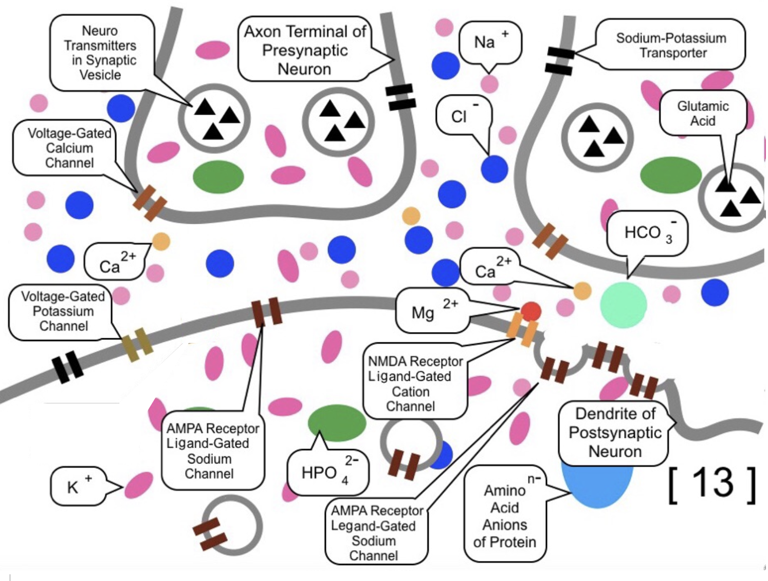
Furthermore, new dendritic spines may be formed in association with this increased receptor density, physically altering the neural structure.
In summary, the enhancement of transmission efficiency at specific synapses—centered around NMDA receptors—constitutes the fundamental biological mechanism of memorization.
*
"Long-Term Potentiation in Wikipedia" https://en.wikipedia.org/wiki/Long-term_potentiation
*
"Short-Term Memory in Wikipedia" https://en.wikipedia.org/wiki/Short-term_memory
*
"Protein Kinase in Wikipedia" https://en.wikipedia.org/wiki/Protein_kinase
2.5.3.2.3 Signal Inhibition through Inhibitory Chemical Synapses
As mentioned above, certain chemical synapses inhibit the transmission of signals from other synapses.
These are called Inhibitory Chemical Synapses. They prevent signal propagation by interfering with the rise of the intracellular electrical potential.
Like excitatory synapses, inhibitory synapses contain neurotransmitters in the axon terminals of the presynaptic neurons. However, inhibitory neurotransmitters differ from excitatory ones like glutamate. Common inhibitory neurotransmitters include GABA (gamma-aminobutyric acid) and glycine.
*
"Gamma-Aminobutyric Acid on Wikipedia"
https://en.wikipedia.org/wiki/Gamma-Aminobutyric_acid
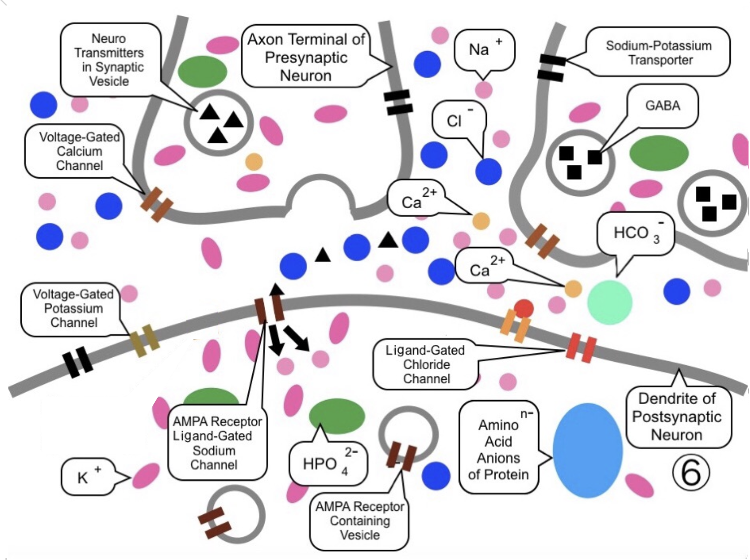
Specifically, as shown in ⑥, the right axon terminal forms an inhibitory synapse coupled with Ligand-Gated Chloride Channels. In this example, the neurotransmitters released by this terminal are GABA. At the same time, the left axon terminal (an excitatory synapse) has released glutamate, causing sodium ions ($\mathrm{Na}^{+}$) to flow in and the electrical potential to rise.
In ⑦, when the positive charge reaches the right axon terminal, calcium ions ($\mathrm{Ca}^{2+}$, in orange) flow in, triggering the release of GABA. The chloride channels receive GABA and open, allowing chloride ions ($\mathrm{Cl}^{-}$, in blue) to flow into the postsynaptic neuron. Because chloride ions are negatively charged, they reduce the intracellular electrical potential (causing hyperpolarization).
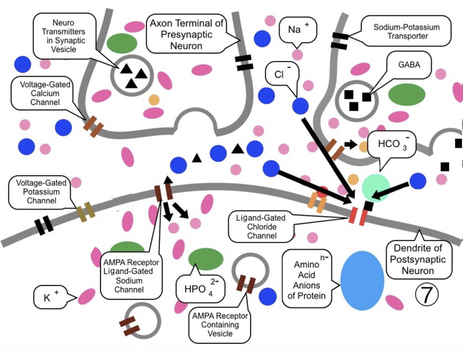
As a result, in ⑨, the rise in potential caused by the left excitatory synapse is
neutralized by the negative influx from the right inhibitory synapse. Therefore, the
postsynaptic neuron fails to reach the threshold and does not fire (send a signal).
Return to the Home Page